
Introduction
Digital pathology
“Eyepiece to monitor”
The 8K imaging System brought to you by SalTus A Life Science Biotech LLC includes 4 key steps:
(1) imaging acquisition (Capture with a 6K camera with your own microscope, 8K in the near future)
(2) process control, storage and management (saving) on high powered work station with a high-end video graphics card.
(3) manipulation and annotation (with editing) say on Adobe Photoshop.
(4) and viewing, display or transmission (sharing) of images on 8K Monitor(s) all in real time.
Notice: High powered workstation with high-end video graphics cards may be necessary to process millions of data points in real time with an 8K system (patent pending)
Digital microscopic 8K images can be used to make preliminary research analysis, consultation, archiving and sharing, education for telepathology, quality assurance of re-review and proficiency testing, image analysis, research and publications, marketing and business purposes presentation.
A mindset of technophobic for microscopic digital pathologist has hindered the adoption of digital pathology because of cost and technical factors.
Cytology slides frequently contain 3D cell groups underneath the coverslip, same plane.
The ability to view these groups in focus on a digital 8K image system can be achieved by multiplane scanning along multiple z-axes or intercalation of scanned images along with different focal points.
The is a disruptive technology, defined as technical innovation that improves a product and/or service in a manner that the market does not anticipate. Computer-aided research analysis of digital images is something more than the traditional microscope can offer, increasingly important as anatomical pathology requires more quantitative image analysis, making more accurate and consistent research analysis with a clear and resolute 8K digital imaging system to monitor from SalTus.
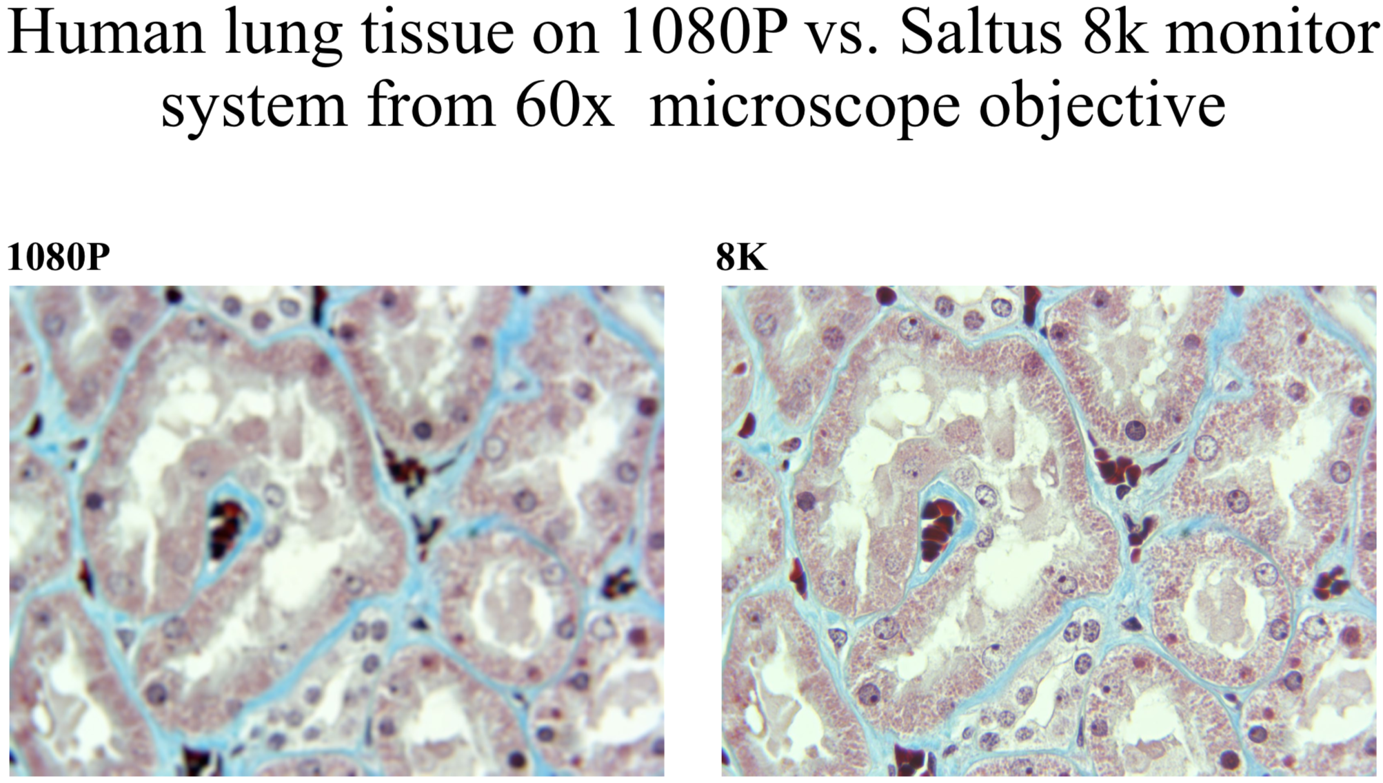
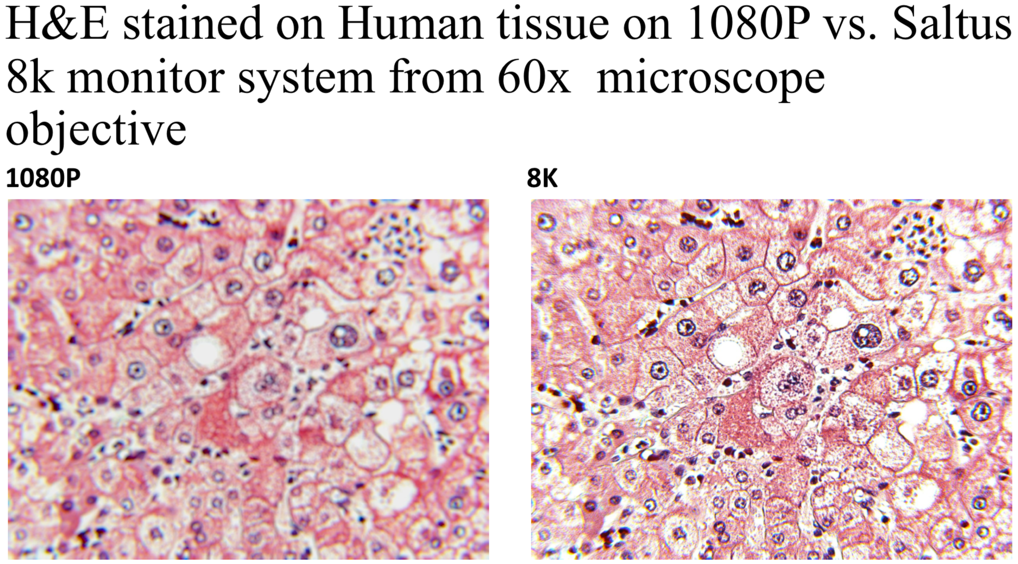
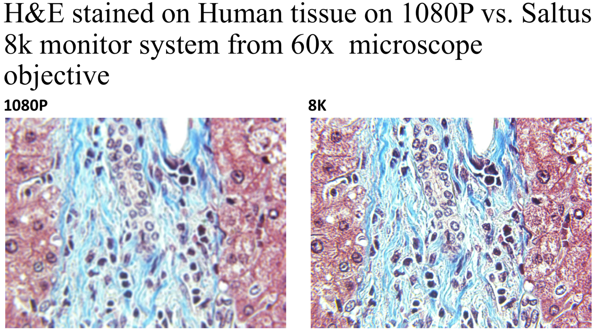
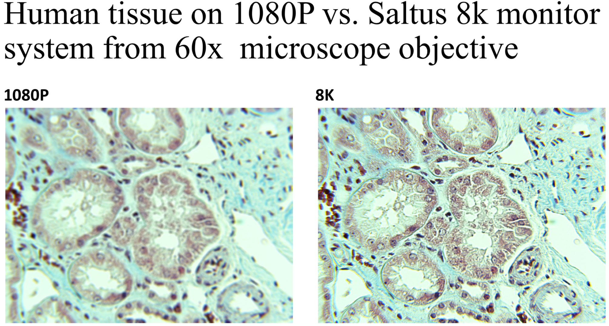
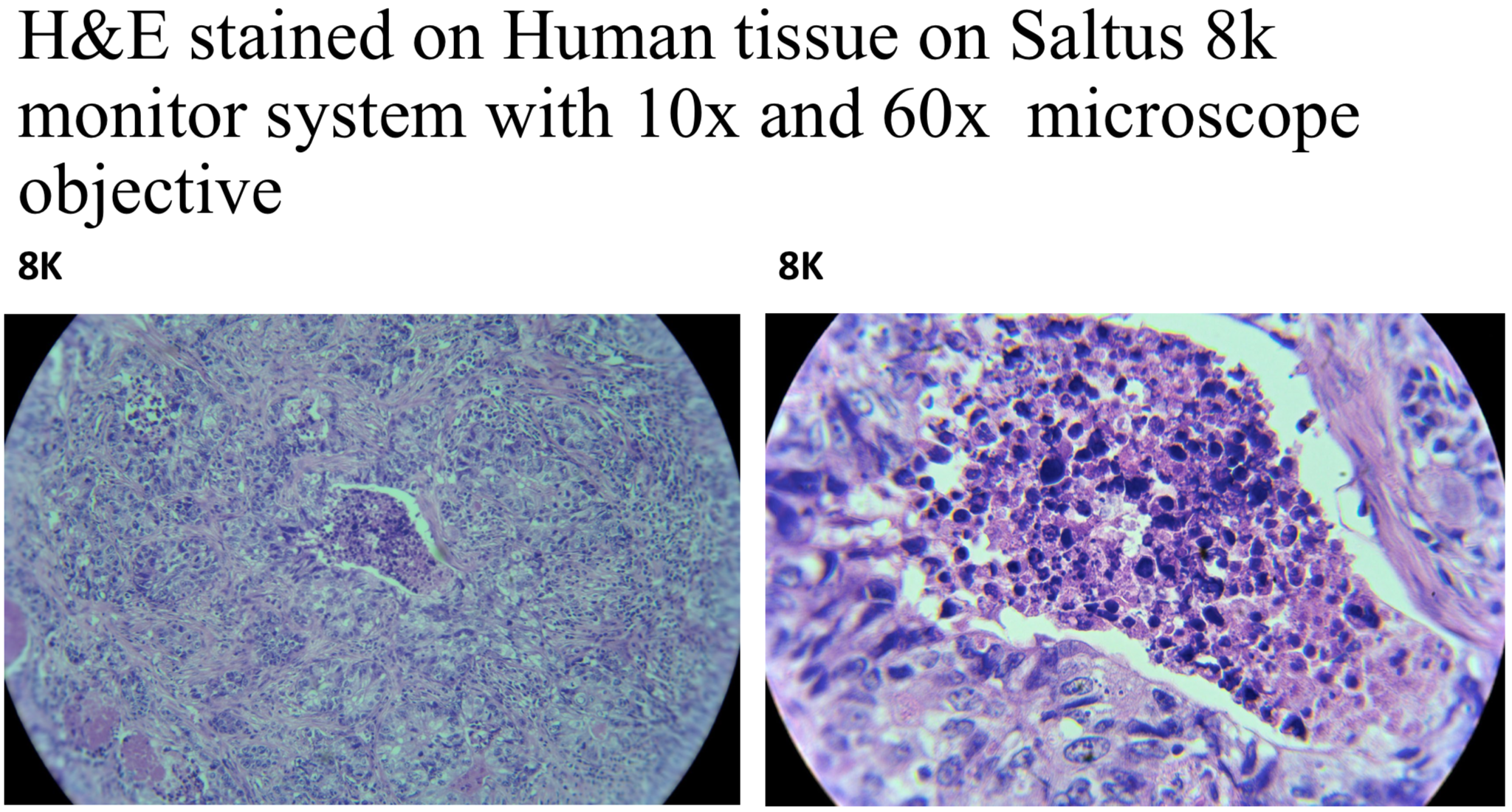
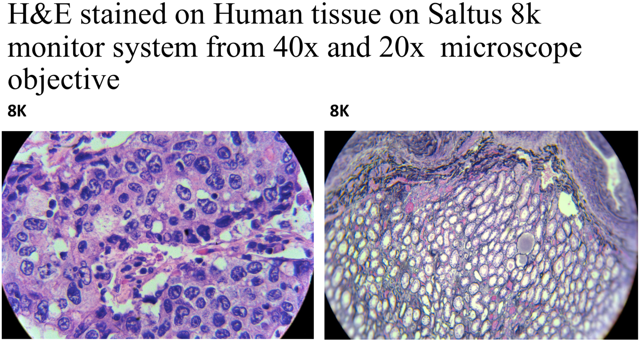
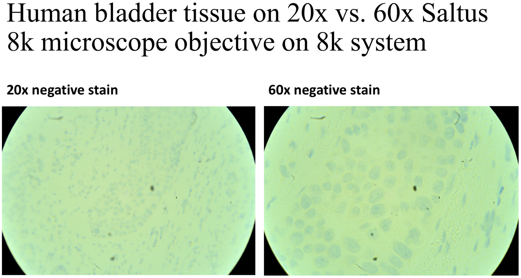
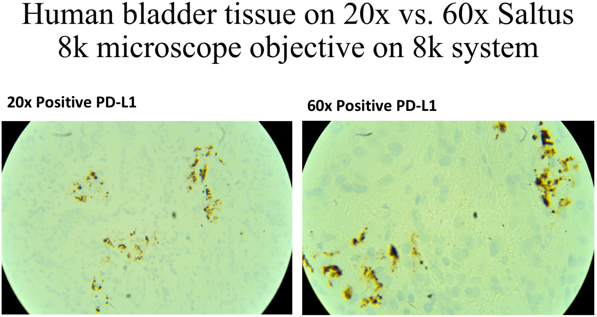
Conclusion: Clarity and Resolution of images are evident on 8K SalTus imaging system
- Capturing digital slides (images) by using 8K system is a necessity with an 8K monitor, every pathologist will want one.
- High-end computer work station provides real-time storing and archiving the images with high definition.
- Editing and performing other manipulations with the captured images saves time, faster and more efficient workflow, cutting cost.
- The detail and higher pixels presentation prevent errors in analysis and adds discovery, integrated with LIS systems.
- Viewing and sharing images with other researchers, doctors or institutions, better outcomes for patients.

Manual Process
Digital Pathology with 8k System
SALTUS BIOTECH
ENABLING PATHOLOGIST TO SEE CELLS BETTER!
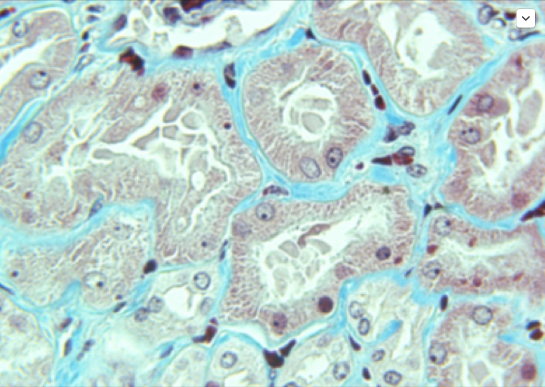
Not Clear Image
1080 P (Today’s old technology)
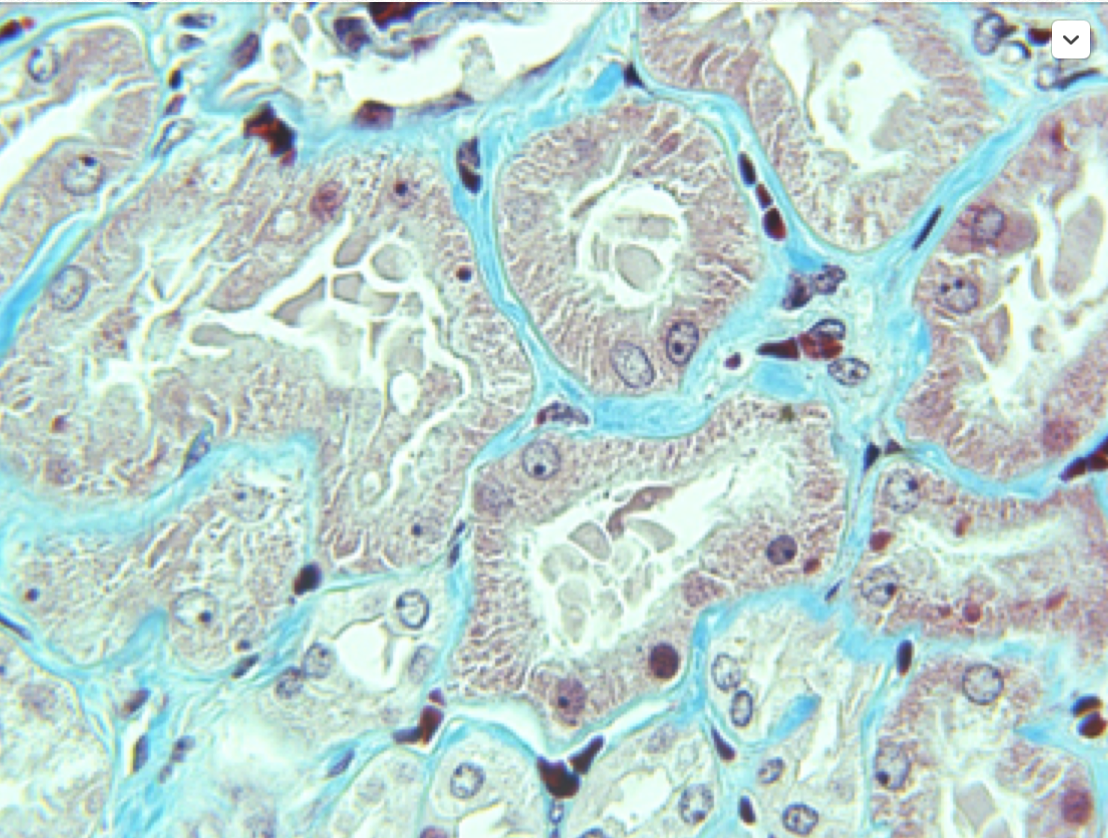
SALTUS Super Clear Image
8K (SALTUS -State of the art)
SALTUS 8k Imaging system dramatically increases clarity and resolution of every biopsy slide.
And the SALTUS 8k imaging system does something no other system in the world does
– Microscopic Movies of live biological tissue specimens. Patent pending
When Pathologists see cells better with SALTUS Biotech 8K Imaging system:
· Ease of use is better!
· Accuracy is better!
· Precision is better!
· Efficiency is better!
· Analysis is better!
· Viewing education slides is better!
· Your Published data is better!
· Cost savings is better!
· Sharing digital data files is better and easy!
Now, Saltus Biotechnology presents state of the art 8k Microscopic Movies TM for Digital Pathology…. As we all know, Digital Pathology will become one of the most important medical technology in the last 10 years for your lab and of the entire healthcare system.
A digital microscope (or imaging system) is an integrated design system that combines a traditional optical light microscope or tri-nodular fluorescent microscope, digital multimedia/8k camera, and digital processing technology with the most powerful workstation you can buy, a digital microscope imaging system typically includes six components: Microscopy, Adaptor, optical module-camera, data acquisition module, digital image processing, and software control modules.
Among these modules, the optical module realizes the function of the microscopic imaging; data acquisition module records the images produced by digital video devices, including CMOS digital camera stored in optical module in the digital format, and then transfers these digital images to the computer storage devices through different graphics card interface or USB interface; and the software control module, the core of the whole system, controls the image capture, processing, and measurement in real-time to optimally improve the image quality, even as microscopic movies. The digital images can be monitored in real-time using an 8k color computer monitor directly from the specimen on the microscope stage.
“Eyepiece to Monitor in Real Time”
We all have seen radiology and other medical disciplines in the healthcare system reaping the benefits of using computer technology in bettering their efficiency and quality. Digital pathology seems like the next in line for efficiently cost saving with Quality data files.
But is that really the case? To find this out, in this presentation we have illustrated the benefits and challenges of adopting 8k digital pathology.
WHAT IS 8K DIGITAL PATHOLOGY, AND HOW DOES IT WORK?
8k Digital pathology stands for using technology in order to speed up and enhance workflow in a pathology laboratory.
In practice, it means using advanced digital 8k microscope systems to capture digital slides, then store files, analyze and share with others.
The entire digital pathology system or environment is comprised of five key components:
· Capturing digital slides (images) FFPE or live tissue by using digital 8k Microscopic Movies TM
· Storing and archiving slides with cells and micro-morphology images of tissues in 8K resolution equates to 7,680 × 4,320, or 33 million pixels (33,117,600, to be exact) on an 8k Monitor with very high-end computer workstations and 8k camera for Microscopic Movies TM from Saltus Biotech
· Editing and other performing manipulations with the captured images and movies
· Viewing and sharing images with other doctors or institutions
All this is done using the Field of View (FOV) to Whole slide images (WSI) technology, or as some call it “virtual microscopy”. In the first phase, “imitate” classic light microscope or fluorescent microscope can capture digital 8k data slides and movies, while in the second phase a specialized software is being used for image viewing and analyzing.
· Last and greatest advance, Live biopsies tissues studied at greater than 24 frames per seconds (fps) movies on 8k system. Viewing PD1/PD-L1 or CTLA-4 checkpoints on live tissue in real time Microscopic Movies TM viewing drug treatment on live tissue specimens.
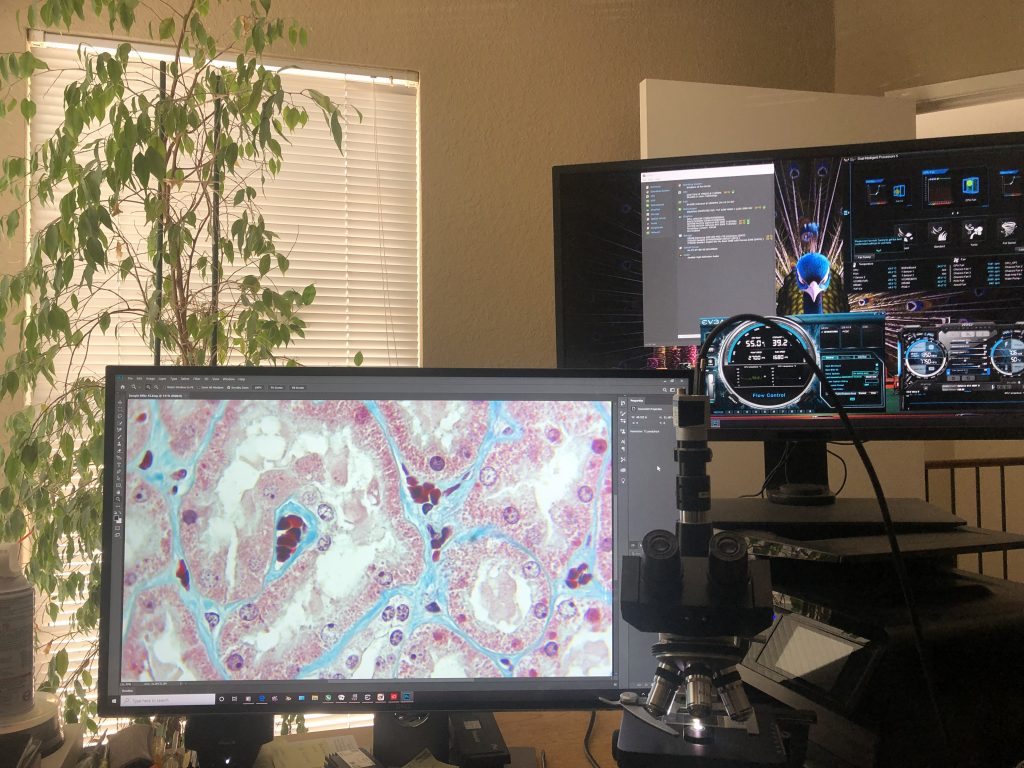
8k Digital System
So, this process replaces the traditional workflow where a pathologist inspects each cut fixed tissue on a glass slide and makes notes and final report conclusions.

WHAT ARE THE BENEFITS?
There are many benefits of implementing an 8k digital pathology system, whether it’s used in laboratories, universities, or research centers.
A digital pathology system, once implemented, will inevitably bring many changes.
These changes are imminent because this is a shift from an analog to a digital working process.
Pathology has witnessed similar changes in radiology some 15 years ago when technology eased the workflow and helped in cutting down the expenses in healthcare.
- Faster and more efficient workflow
The advantage of digital pathology is using digital 8k system with Microscopic Movies TM
and computer software to process, analyze and share digital images of tissues specimens electronically with doctors or entire teams of doctors around the world.
Once the image of the tissue is captured, it can be used an indefinite amount of times for data results, sharing, and educational purposes. It’s clear that transporting glasses is a delicate matter due to its fragile nature.
The digital image can also be used for pathological reports, as well as for result research prognosis by using advanced specialized computer software for image analysis.
- Cutting costs
In a period of 5 years after implementing digital pathology, expected savings are estimated at 10’s million dollars for a laboratory that had several 100 thousand orders per year.
One of the key factors is better time management by laboratory staff due to increased productivity.
Efficiency and precision of digital pathology system in analyzing tissue images is much higher than the one of a classic light microscope manual process.
- Better outcomes for patient’s clear resolution and clarity in 8k Digital microscope systems
Along with cost savings, better outcomes for patients will be noticed with much more accuracy and precision leading to less incorrect analysis. Each day, more patients are asking their pathologists whether they feel confident about their analysis results. This puts pressure on pathologists to deliver precise data in a timely manner, and digital pathology can help pathologists achieve better results.
Present day, with the abundance of patient data available to clinical doctors, it’s difficult to deliver a fast and accurate analysis data without the help of our 8k system technology. 8k Digital pathology can make a difference, as it automatizes and speeds up routine tasks which would be impossible for a pathologist to do in a short period of time without compromising quality.
As soon as a pathologist receives the biopsy images, they can forward digital files via a link to their colleagues anywhere in the world.
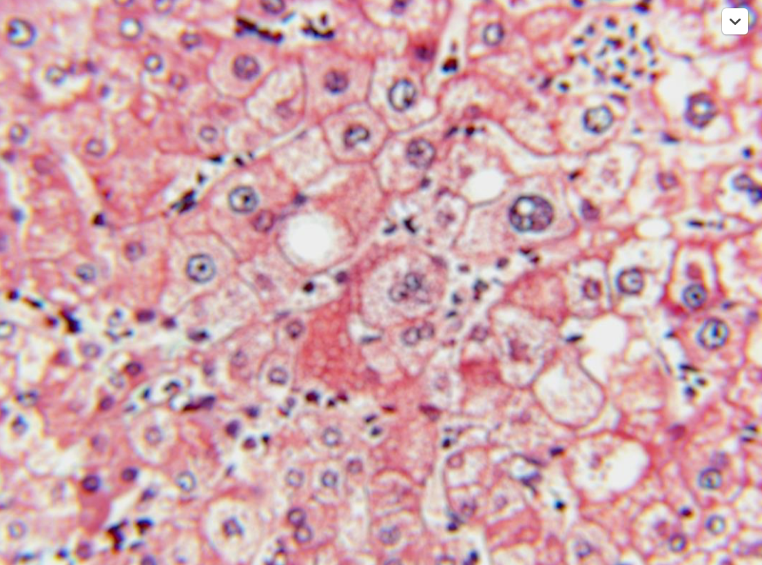
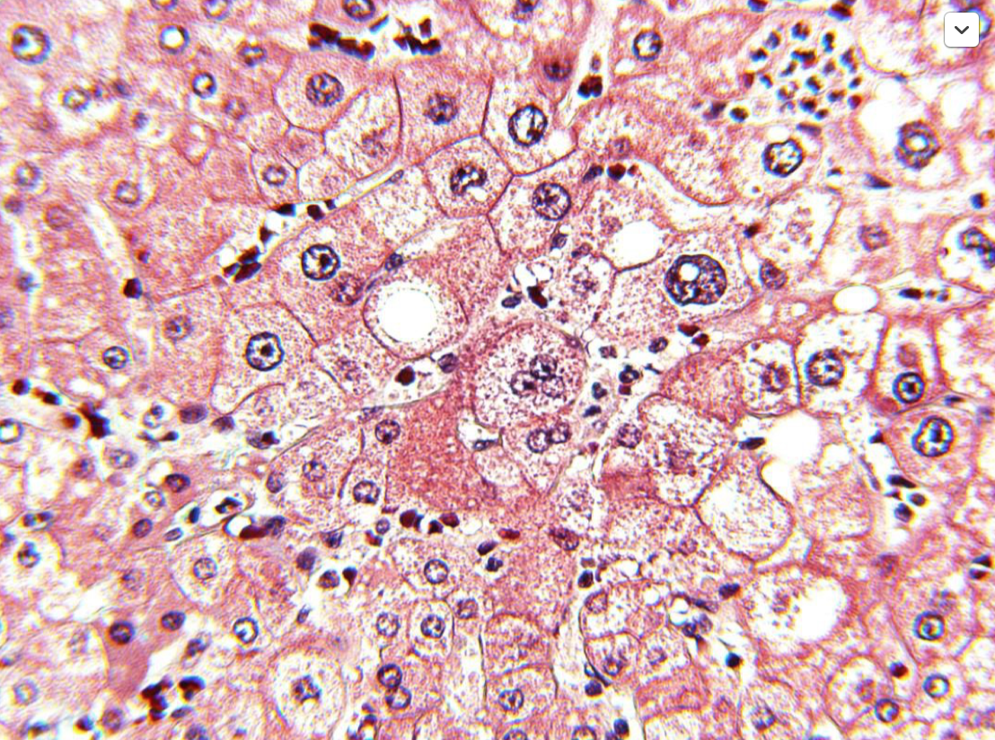
H&E stained on Human tissue on 1080P vs. Saltus 8k monitor system from 60x microscope objective
1080p resolution
8k clearer resolution
- Digital images integrated with LIS systems
8k System Technology will be finding increasing roles in every part of the healthcare system. Laboratory informational systems are implemented in most half-decent laboratory, and we can say that LIS is already old news.
In institutions where hospital and laboratory information systems are integrated, classic analog workflow makes the pathology laboratory seem like a slow ancient process with no real connection to other hospital departments.
But, when a Saltus 8k digital pathology system is installed, it will make sharing digital images an easy task, thus connecting pathology laboratory with the hospital departments and the entire healthcare system.
Integration of these technologies into a single powerful 8k digital system is a great platform for creating better multidisciplinary expert teams, organizing virtual meetings, publishing data as well as for educating new experts.
Digital pathology in 8k systems represents more of an evolution than a revolution in healthcare. And with all these benefits you must be asking yourself: “Why isn’t digital pathology implemented everywhere as we speak?”
Well, the answer to that question isn’t easy, as there are many obstacles.
These problems, or let’s say challenges, demand to be addressed. Saltus will help you dive into it in our next generation of Digital Pathology.
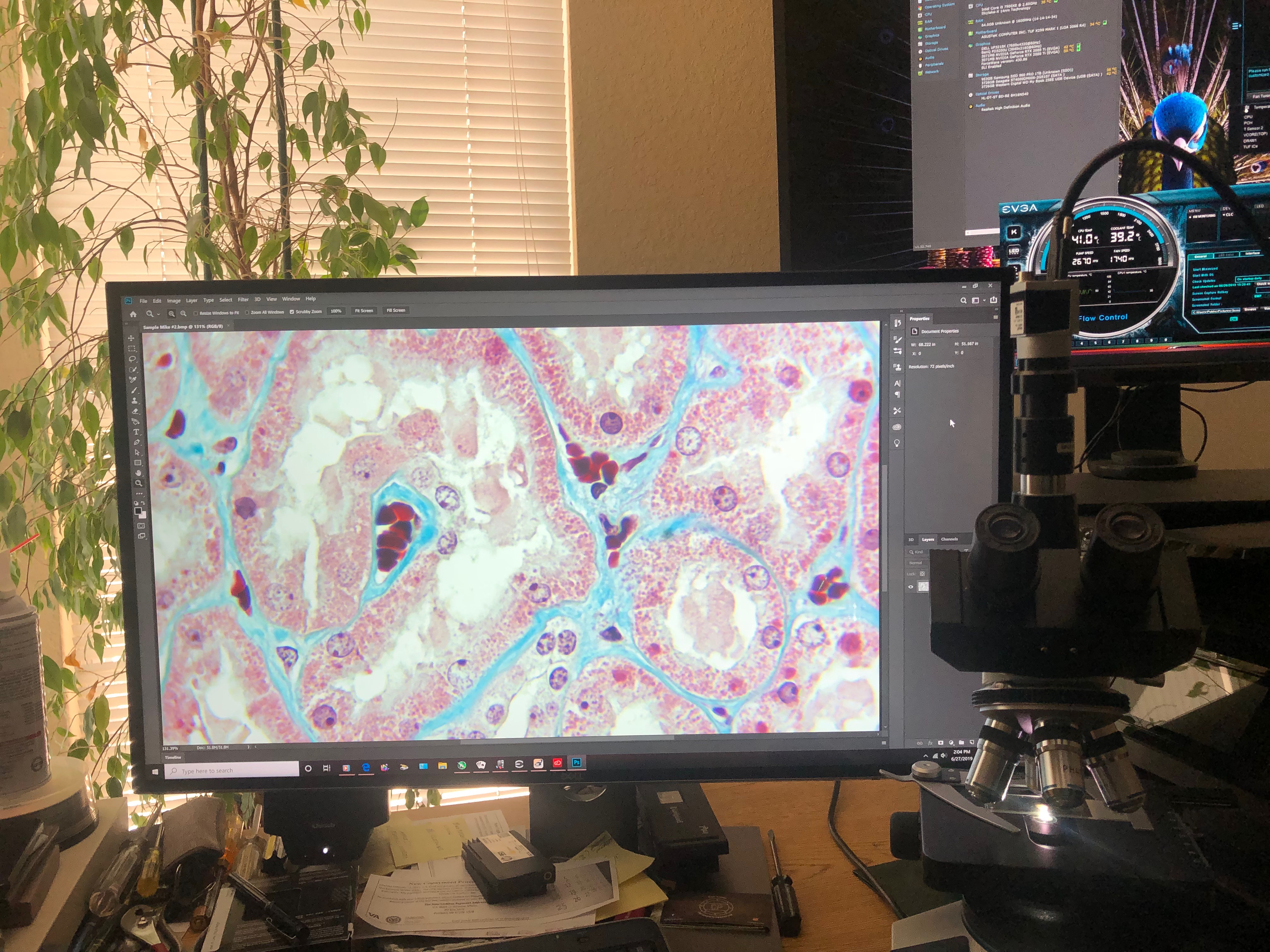
8k Monitor Digital Image System
CHALLENGES FOR DIGITAL PATHOLOGY
Process of adopting new technology is never easy, Saltus understand this.
There are issues to tackle in order to improve pathology.
· Technology Fear
Even though fear may be a bit aggressive, we have all had some experience with it in everyday life.
Adoption of new 8k computer technologies and trusting them is a big challenge for some people, but that’s ok. It is normal to just not feeling comfortable using them.
“Change is always difficult. we think, while we’re looking through our microscopes, we just feel safe because that’s what he learned through years of practice.”
In 8k Digital pathology, years of working by following standard process and workflow, in a familiar environment result in developing habits and “comfort zones”.
And truly, pathology is conservative and relies on previously acquired, proven knowledge.
Viewing a tissue sample on a monitor screen instead of a microscope is a huge change.
In this new digital environment, pathologists need to get familiar with Whole Slide Imaging (WSI) which implies acquiring excellent skills in using digital scanners/microscopes as well as the specialized software for image analysis and manipulation.
This asks for additional education and adaptation.
Luckily, this adaptation will be much easier for the new generation of young pathologists who are used to using technology in every situation.
· Possible Challenges
There are few issues which cause caution and concerns about 8k digital pathology, and they all come back to technology. One of the most important factors in implementing 8k digital pathology is computer hardware a very powerful workstation.
For example, if you use a monitor with bad color accuracy it can lead to inaccurate results.
We have mentioned 8k monitors as an example, but in reality, it is just one piece of the puzzle needed for completing a successful 8k digital pathology system.
It’s very important that the 8k pathology” workstation” used for viewing and manipulating digital images has the best, most up to date computer hardware. Also, capturing high-resolution images creates a huge amount of new data 33 million color pixels per second. This demands investment in increasing the existing storage capacities.
An average laboratory can make 300 to 500 new slides daily, which means additional 1000 GB or more of data each day.
Additionally, having a LIS system implemented will increase the overall data transfer speeds which will result in faster processing — all the way from capturing an image up to the image appearing on the doctor’s “desk” or lab top while on the road.
DIGITAL PATHOLOGY MARKET
How big is Saltus 8k digital pathology market? And is growing each day.
Estimated market growth for digital pathology systems in Europe is huge, and it will increase from 62.23 million dollars in sales (2012) to 143 million dollars in 2019.
In the USA this growth is even bigger — 205 million dollars in 2019.”

It’s worth pointing out that in 2011 we had somewhere in between 1000 and 1500 digital pathology systems implemented in institutions around the world, which is still a very small adoption percentage.
But still, these numbers are getting bigger each day.
WRAP-UP
Digital pathology is a disruptive 8k technology and innovation changing the core of processes in pathology.
DP will enable better primary biopsies results, helping pathologists easily access digital images or Microscopic Movies from different sites, and create a platform for better multidisciplinary teams.
Also, this 8k technology has a huge potential for educating new generations of pathologists.
Standardization and regulation of digital pathology is not at the point where it should be, and pathologists still have certain issues with adopting this new 8k technology.
And we can’t ignore the fact that these digital pathology 8k systems will pay for themselves in a short period of time. But as the market grows, these systems will be much more of a necessity for medical institutions around the world.
It seems that the adoption of 8k digital pathology systems will be an imminent process.
So, Contact Saltus Today and be the First with 8k Digital System!
SalTus Biotech LLC
PO Box 12601
Pleasanton, Ca. 94588
Webpage: SaltusBioTech.com
For Research Use Only, Not for Diagnostics
7/2019
